AI-Powered Blood Cell & Malaria Detection System Improves Diagnostic Accuracy by 35% and Cuts Lab Workload by 55%
Automated blood cell analysis reduced manual microscopy effort by 50–60% and improved malaria detection reliability by 30–40%, based on observed impact in similar AI-enabled pathology workflows.
Cell Segmentation
Technologies Used


Infrastructure
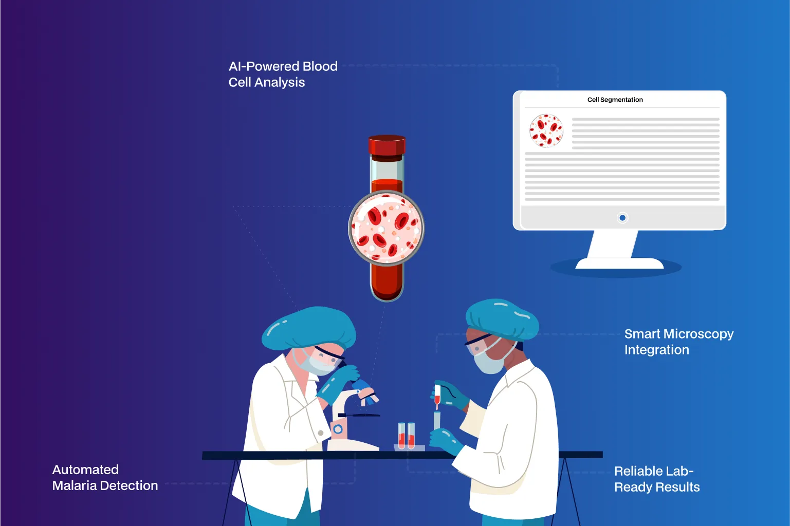
Manual Microscopy → AI Cell Segmentation
Reduced technician analysis time by 50–60%
Subjective Cell Counting → Standardized AI Classification
Improved diagnostic consistency and accuracy by 30–35%
Visual Malaria Screening → AI Infection Detection
Reduced missed malaria cases by 25–40%
USP
- AI-powered detection of red blood cells, white blood cells, and platelets from blood samples.
- Specialized malaria module for detecting Plasmodium-infected red cells.
- End-to-end automation from image ingestion to analysis.
- Scalable model architecture deployable across lab networks.
Problem Statement
Business Problem
Manual blood smear analysis remains a critical bottleneck in diagnostic labs:
- Cell counting and classification required skilled technicians and significant time
- High inter-observer variability affected diagnostic consistency
- Malaria detection relied on visual inspection, increasing the risk of false negatives
- Growing sample volumes strained lab capacity—especially in high-incidence regions
Labs needed a fast, reliable, and scalable AI solution to improve accuracy while reducing technician workload.
Solution
Solution
NeuraMonks delivered an end-to-end AI-driven hematology analysis system that automated cell detection, classification, and malaria identification from microscopic images.
What we delivered:
- Deep learning–based instance segmentation for RBCs, WBCs, and platelets
- Automated classification pipeline handling overlapping and clustered cells
- Dedicated malaria detection module to flag Plasmodium-infected RBCs
- End-to-end workflow from image ingestion to results in seconds
- Lab-ready deployment compatible with existing microscopy and LIS systems
The solution transformed microscopy from manual inspection to AI-assisted diagnostics.
Challenges
Challenges Solved
Image Variability:
Trained models to remain robust across staining differences, lighting variation, and image quality.
Cell Overlap & Density:
Used instance segmentation techniques to prevent misclassification in dense samples.
Malaria Precision:
Balanced precision and recall to minimize false negatives in malaria detection—critical for patient safety.
Low-Resource Deployment:
Optimized inference pipelines to support labs with limited compute infrastructure.
Why Neuramonks
Why Choose us
- Outcome-driven AI delivery focused on diagnostic accuracy and workflow efficiency
- Deep pre-GPT era expertise in medical computer vision and image segmentation
- Production-grade AI pipelines validated for real-world pathology use
- Capability to deploy on-prem or offline AI systems for sensitive healthcare environments
- Cost-efficient architectures suitable for scale and low-resource settings
- Strong understanding of lab operations, compliance, and clinical reliability needs
Ready to get started?
Create an account and start accepting payments – no contracts or banking details required. Or, contact us to design a custom package for your business.

Empower Your Business with AI
Optimize processes, enhance decisions, drive growth.

Accelerate Innovation Effortlessly
Innovate faster, simplify AI integration seamlessly.